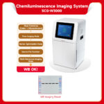
SCG-W3000 Chemiluminescence Imaging System
SCG-W3000 chemiluminescence imaging system is equipped with a high-sensitivity camera with 9 million pixels. It enables fast, accurate, and high-throughput detection and imaging of samples, and is widely used in fields such as life sciences, medicine, and environmental protection.
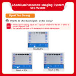
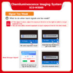
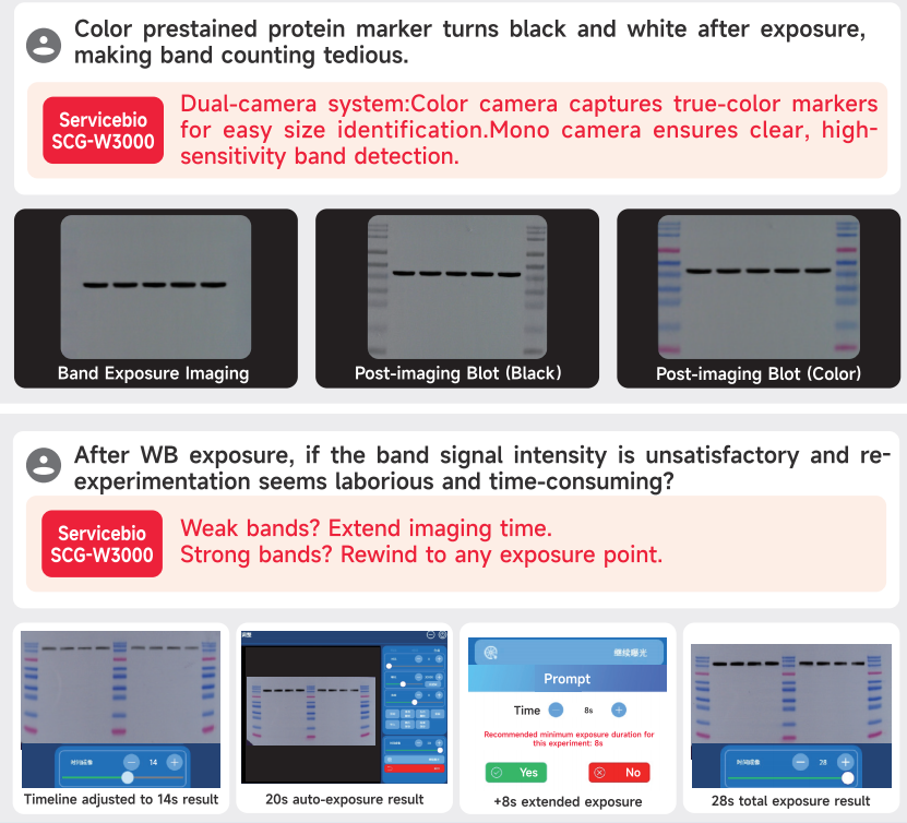
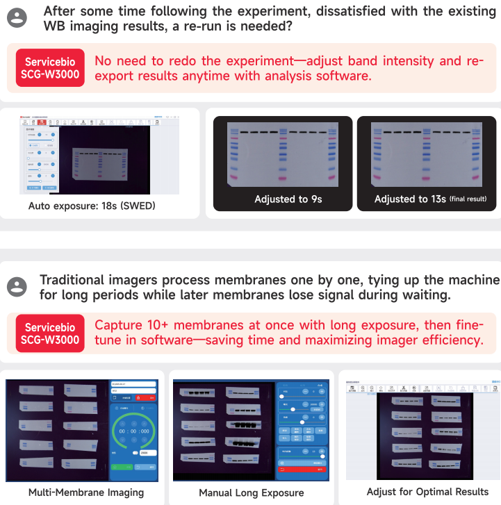
- Whether the signal is weak or strong, a single imaging session can deliver your WB results.
- For weak signals, you can directly extend the imaging time based on the original image for further imaging.
- For strong signals, you can adjust the timeline to retrieve any result image from the imaging duration.
- Supports simultaneous imaging of up to 10 membranes (or even more), improving instrument efficiency—1 device equals 10!
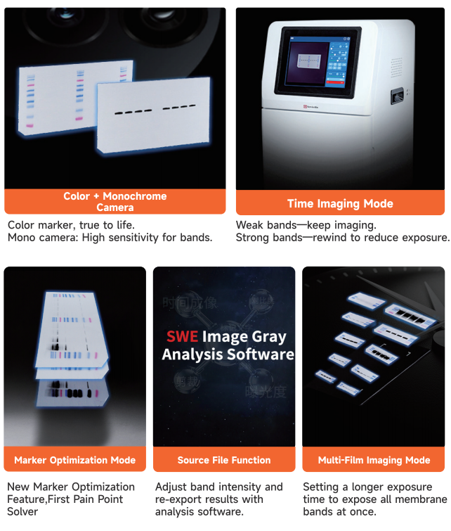
- Dual-camera system:
- A color camera captures markers with true-to-life colors for greater authenticity.
- A monochrome camera captures bands with high sensitivity, delivering superior performance for weak signals.
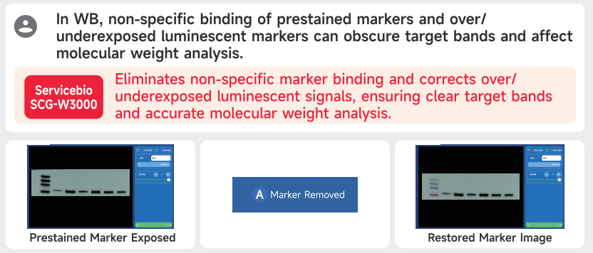
- Marker optimization function:
- Eliminates nonspecific band signals caused by prestained markers and antibody interactions.
- Adjusts overexposed or underexposed chemiluminescent marker signals.
- Source file storage allows flexible adjustments anytime:
- If bands appear too thick or too thin after the experiment, you can easily fine-tune the results on your computer using the source file function!
Working with the ECL Chemiluminescence Kit (G2161-200ML), it meets the exposure needs of high, medium, and low-concentration samples. Enjoy a better WB imaging process and achieve superior WB imaging results!
SWE Imaging Grayscale Analysis Software-SCG-W2000 SCG-W3000-3.3.2-win32-x64-setup.exe
Download link from Google Drive: https://drive.google.com/file/d/1IoXazML9PDHoN4HqOYm-t3C2bVFvQ_rt/view?usp=drive_link
SOFTWARE USER MANUAL:
SWE Imaging Grayscale Analysis Software-SCG-W2000 SCG-W3000.pdf
| Product Name | Chemiluminescence Imaging System | |
| Cat. No. | SCG-W3000 | |
| Dimensions | 400mm×371mm×700mm | |
| Camera 1 | Pixel Resolution | 9 million pixels |
| Resolution | 2992*3000 | |
| Pixel Size | 3.76μm×3.76μm | |
| Target Size | 1″ (11.28mm x 11.28mm) | |
| Full Well Capacity | 16.5ke-(HCG),50.5ke-(LCG) | |
| Sensitivity | 877mv@1/30s | |
| Readout Noise | 1.24e-(HCG), 3.22e-(LCG) | |
| Dark Current | 0.0003e-/s/pixel@-15℃ | |
| Signal-to-Noise Ratio | 42.2dB(HCG), 47dB(LCG) | |
| Exposure Time | 0.1ms~1h | |
| Binning Mode | 1×1, 2×2, 3×3 | |
| Grayscale | 16-bit (65536 levels) | |
| Cooling | Relative to Ambient Temperature-40℃ | |
| Camera Type | Black and White Camera | |
| Camera 2 | Pixel Resolution | 45 million pixels |
| Resolution | 2992*3000 | |
| Pixel Size | 2.315×2.315μm | |
| Target Size | 1.4″ (18.93 × 13 mm) | |
| Full Well Capacity | 10.8ke- | |
| Sensitivity | 419mv | |
| Readout Noise | 2.12e- | |
| Dark Current | 0.12mV | |
| Signal-to-Noise Ratio | 40.3dB | |
| Exposure Time | 17μs~15s | |
| Binning Mode | 1×1, 2×2, 3×3 | |
| Grayscale | 8 bits (256 gray) | |
| Camera Type | Color Camera | |
| Lens1 | Aperture | F0.95-F16 |
| Focal Length | 17mm | |
| Type | Prime lens | |
| Light Source | Bright Field Light Source | Downward-facing LED white light source, symmetrically distributed on both sides |
| Dark Box | Light Isolation | Fully light-sealed, isolates environmental light. |
| Door Control | Door control sensor can control the on/off of the bright field light source. | |
| Field of View | Effective field of view is 136mm*136mm (expandable to 200mm×200mm if needed). | |
| Auto Exposure | Intelligent exposure technology can quickly determine the optima exposure time and automatically perform binning. Combined with time-lapse imaging and time accumulation functions, users can achieve the bestimage results with just one opematchration. |
|
| Software Functions | Exposure Modes | High Quality:Highest image quality Standard:Balancesimage qualityand exposurespeed High Sensitivity:Fastestexposure speed |
| Real-time imaging | Real-time presentation of the changes in sample signals during the exposure process, allowing for the observation of every detail of the capture. Overexposed areas will be indicated for samples with overexposure. |
|
| Time Imaging | After exposure is complete, each frame image within the exposure time can be generated.Through precise retrospective adjustments users can choose any frame image within that exposure time as the final output. |
|
| Time Accumulation | For samples with insufficient exposure, users can choose to continue exposure after the initial exposure is completed, enabling the sample to receive additional exposure on top of the already exposed time. |
|
| Optional settings | Color Marker/Black & White Marker, Overexposure Prompt/No OverexposurePrompt |
|
| Industrial Computer | 10.4-inch display with a resolution of 1024×768, running on Windows 10 operating system, featuring 16GB of RAM, 512GBssD, and built-in Bluetooth and WiFi. |
|
| External Interfaces | 2 USB 3.0 ports | |
| Operating Voltage | 90~132VAC/180~264VAC (selectable via switch), 47~63Hz. | |
| Product Power | ≤200W | |
| Product Net Weight | 23.45Kg | |
Notes:
- Do not touch or scratch the internal lens of the dark box with hands or sharp objects.
- After placing the sample, ensure the instrument door is properly closed to prevent external light from entering and affecting results.
- During imaging, avoid shaking the workbench or instrument to prevent compromised image quality.
- Observe electrical safety—do not pull the power cord or move the instrument during operation.
- After experiments, clean the dark box thoroughly, removing all samples and residues.
Parameter Comparison
| Cat. No. | SCG-W5000 | SCG-W3000 | SCG-W2000 |
| Dimension | 400×371×700 mm | 400×371×700 mm | 400×371×700 mm |
| Camera | Depth-cooled high sensitivity camera | Depth-cooled high sensitivity camera | High-sensitivity camera |
| Resolution | 2992×3000, 9 megapixels | 2992×3000,9 megapixels +2992×3000
45 megapixels |
3072×2048, 6.3 megapixels |
| Pixel | 3.76×3.76 μm | 3.76×3.76 μm | 2.4×2.4 μm |
| Shooting Area | Effective field of view for blotting film/protein gel: 136×136 mm Effective field of view for nucleic acid gel: 140×140 mm. |
Blotting Film 136×136 mm |
Nucleic Acid Gel/Protein Gel 140×140 mm |
| Cooling Temperature | Relative ambient temperature -40°C | Relative ambient temperature -40°C | – |
| Light Source | Bright-field Light Source: Downward-facing LED white light source, symmetrically distributed on both sides. UV Light Source: 310 nm LED array for uniform transmission illumination. |
Downward-facing LED white light, symmetrically distributed on both sides | Bright-field Light Source: Downward-facing LED white light source, symmetrically distributed on both sides. UV Light Source: 310 nm LED array for uniform transmission illumination. |
| Industrial Computer | 1match0.4 inches, 1024×768 Windows operating system |
10.4 inches, 1024×768 Windows operating system |
10.4 inches, 1024×768 Windows operating system |
| External Interface | 2 USB3.0 | 2 USB3.0 | 2 USB3.0 |
| Working Voltage | 90~132VAC/180~264VAC (Selectable via switch)47~63HZ |
90-132V/180-264V | 90~132VAC/180~264VAC (Selectable via switch)47~63HZ |
| Product Power | 200 W | ≤200 W | 200 W |
| Net Weight | 25 kg | 23.45 kg | 30 kg |
| Real-Time Imaging | Yes | Yes | – |
| Time Imaging | Yes | Yes | – |
| Time Accumulation | Yes | Yes | – |
| Auto Exposure | Yes | Yes | Yes |
| Choice of 3 Imaging Modes | Yes | Yes | – |
| Protein Gel/Nucleic Acid Gel Imaging | Yes | – | Yes |
| Nucleic Acid Gel Cutting | Yes | – | Yes |
Literature Citations:
- Wang G., et al. An injectable decellularized extracellular matrix hydrogel with cortical neuron-derived exosomes enhances tissue repair following traumatic spinal cord injury. Mater Today Bio 28, 101250 (2024). PMID 39318371 IF 8.2 (PMID:39318371)
- Liu, S., et al. PTGES3 proteolysis using the liposomal peptide-PROTAC approach. Biol Direct 19, 144 (2024). PMID 39726032 IF 5.7 (PMID:39726032)
- Huang, K., et al. The Role of TPM3 in Protecting Cardiomyocyte from Hypoxia-Induced Injury via Cytoskeleton Stabilization. Int J Mol Sci 25, 25 (2024). PMID 38928503 IF 5.
- Zhang, PMID:40427372
- Tan, C., et al. Lycopene inhibits pyroptosis of endothelial progenitor cells induced by ox-LDL through the AMPK/mTOR/NLRP3 pathway. Open Med (Wars) 2024, 20240973 (2024). PMID 38919547 Application: WB IF 2.1